-
Call us now
081053 75555 -
Mail Us
vardhanfertility@gmail.com -
Mon - Sunday
8:30am to 8:30pm

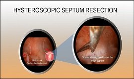
Hysteroscopy and laparoscopy is still the most dependable technique for the diagnosis of uterine septum. With a shorter operative time, less blood loss, a significantly increased postoperative pregnancy rate and live birth rate, and a significantly lower spontaneous abortion rate, transcervical resection of the septum is the preferred method for the treatment of uterine septum, and surgical instruments and skills were critical to the prognosis of uterine septum. Gynecological examination, hysterosalpingography (HSG), magnetic resonance imaging (MRI) and combined hysteroscopy and laparoscopy are used as methods of diagnosis. Before operation, systemic physical examination is performed to exclude surgical contraindications. After anesthesia is administered successfully, an electro resectoscope is placed to confirm the location, size, and range of the septum. The septum is then incised by needle electrode and laparoscopy is used for overall monitoring during the operation. For patients with complications such as infertility, uterine fibroid, ovarian tumor, pelvic adhesions and tubal atresia, additional procedures such as hysteromyomectomy, ovarian tumor removal and pelvic endometriosis surgery were also performed. At the end of the operation, a balloon (filled with 5–8 mL of normal saline solution) will be placed into the uterine cavity.

Join Us! Never go off from any of our updates related to pregnancy, infertility and healthy tips from Vardhan Fertility. Let’s get connected!